You are here

Cord Blood Telomere Length in Latino Infants
Longer Telomeres found in Female Infants & Infants whose Mothers have Higher Education
Background
Telomeres are the tips of the chromosome that prevent degradation and damage. Telomeres are associated with biological aging because with each cell division, they divide and get shorter, so that elderly adults have the shortest telomeres. Telomeres also get shorter with exposure to chronic disease and stress, as they are very sensitive to damage from reactive oxygen species (ROS).
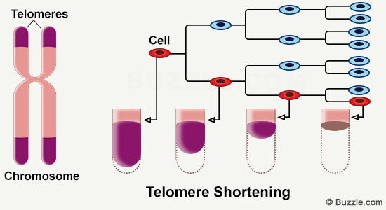
The greatest period of telomere shortening occurs during the first 4 years of life. Only a few risk factors for accelerated shortening have been identified because there have been few studies of telomere length conducted with infants and young children. Some in utero factors have been found to be associated shortening, including exposure to maternal smoking, stress in pregnancy, and babies that are large for gestational age. Yet few studies have evaluated risk factors for shorter telomere length at birth.
Studies of telomere length at birth are needed so that we can better understand risk factors for development of chronic disease, particularly in high-risk populations. In adults, shorter telomere length has been associated with development of diabetes mellitus1 and progression of metabolic syndrome2.
Study Methods
We recruited a cohort of 97 pregnant, self-identified Latina women from a public hospital in San Francisco. Exclusion criteria were all women who had insulin-treated diabetes mellitus, drug or alcohol abuse, or any contraindications for breastfeeding.
Cord blood was successfully collected at delivery from 54 of the study participants. Telomere length was determined from the whole cord blood by quantitative PCR at our laboratory at the University of San Francisco (E Blackburn). We assessed infant anthropometrics at birth (weight, length, head circumference) and maternal pregnancy medical history, including gestational diabetes mellitus, hypertension and weight gain. Maternal socio-demographics were also collected, including education history, marital status, and country of origin.
Telomere length (measured in base pairs) was evaluated in relationship to other infant and child characteristics using t-tests for dichotomous variables and analysis of variance for continuous ones. Multivariable linear regression was used to assess the relationship between telomere length and predictors that were significant at p<0.05 in bivariate analysis.
Results
This was the first time that infant telemere length was studied in a population that was very homogeneous and representative of low income Latinos. All of the mothers were from Mexico or Latin America, and 93% were participants in the Women, Infants and Children’s Program (WIC) which requires low income status.
We found3 that higher maternal education (having a high school diploma or more) was associated with increased telomere length in these newborns. The fact that Latina women with at least a high school diploma were more likely to have babies with longer telomere length suggests that education may be a good marker for exposures to oxidative stress. The correlation between telomere length and education had P value <0.01 and could not be explained by other variations in this cohort, such as mothers with/without prenatal depression, etc.
We also found longer telomere length in female versus male children, suggesting that there may be differences from birth in terms of risk for chronic disease and overall longevity. Studies in adults have similarly found that women on average have significantly longer telomeres than men, but it has been hypothesized that longer telomeres in women may be due to hormonal factors or psychosocial factors which result in longer lives for women. Ours is the first study to find that females have longer telomeres at birth. Further studies are needed to find in utero exposures that may differentially impact female versus male babies.
Latino children are known to be at increased risk for obesity and diabetes mellitus compared with Caucasian children. Our study is the first to assess telomere length in newborn Latino children. Our finding that Latino children already have shorter telomeres at birth if their mothers did not minimally have a high school education suggests that children may be impacted at the cellular level by maternal access to education. These findings have important policy implications as they suggest a need for increased access for GED and high school equivalency programs for immigrant and other high-risk families.
 Janet Wojcicki, PhD, MPH, is an Assistant Professor of Pediatrics at the University of California in San Francisco. Dr. Wojcicki is an anthropologist and epidemiologist with an interest in early life risk factors for the development of obesity in high-risk populations. Specifically, she is interested in maternal exposures in pregnancy and early-life feeding decisions that can increase risk for obesity by age five. Additionally, Dr. Wojcicki is also interested in how pediatric under-nutrition can contribute to HIV progression and infection in sub-Saharan African populations. She is a collaborator on a long-term cohort study in Lusaka, Zambia.
References
- Willeit P, Raschenberg J, Heydon EE, Tsimikas S, Haun M, Mayr A, Weger S, Witzum JL et al. Leucocyte Telomere Length and Risk of Diabetes Mellitus: New Prospective Cohort Study and Literature-Based Meta-Analysis. PLOS One November 2104; 9(11): e112483. doi:10.1371/journal.pone.0112483
- Revesz D, Milaneschi Y, Verhoeven JE and BWJH Penninx. Telomere length as a marker of cellular ageing is associated with prevalence and progression of metabolic syndrome. J Clin Endocrinol Metab 2014a Dec; 99(12): 4607-15. doi:10.1210/jc.2014-1851
- Wojcicki, JM, Olveda, R, Heyman MB, Elwan D, Lin J, Blackburn E, Epel E. Cord blood telomere length in Latino infants: relation with maternal education and infant sex. J Perinatology December 2015; doi:10.1038/jp.2015.178