Вы здесь


Buyer Beware: Parents should demand quality testing of umbilical cord tissue storage
Background: cellular therapies from cord blood and cord tissue
Stem cells promise a tremendous potential for treating patients with cellular therapies. The stem cell sources from bone marrow, peripheral blood, and cord blood have been used for at least three decades in over 80 therapies approved by the FDA1. They are now regularly used for treating patients globally.
In recent years we have been exploring new sources for cellular therapy and one such source is the Mesenchymal Stromal Cells (MSC) in umbilical cord tissue. The umbilical cord is an extension of the placenta and the membrane lining the cord is part of the amniotic membrane covering the placenta. Many researchers feel that perinatal tissues are the best source for harvesting MSC, because their collection is noninvasive and their proliferation and immunomodulatory properties seem to be better when compared to adult sources2.
The clinical applications of MSC’s are promising. Between 2011 and 2016 more than 700 clinical trials with MSC were registered in international databases3. Almost 100 papers have been published about human studies using umbilical cord MSCs4. The website of Parent’s Guide to Cord Blood Foundation has a search portal that displays all currently recruiting clinical trials with MSC from umbilical cord tissue. Some of the most exciting applications of MSC are for auto-immune disorders, heart disease, and orthopedics5-10.
Steps of umbilical cord tissue banking
Below we explain each of the steps of umbilical cord tissue banking. Readers who want to know the key take-aways about quality testing can jump ahead to that section.
- Maternal History
- Collection
- Transport
- Processing
- Quality Testing
- Cryopreservation
 Maternal History questionnaire is important for any perinatal blood or tissue banked for therapeutic use.
Collection: After the baby has been delivered, the umbilical cord is clamped and cut. The cord is cleaned with a disinfectant. Once cord blood is collected from the umbilical cord, the cord itself can be harvested for tissue. Usually a section of the cord measuring 2-4 inches or 8-10 cm in length is cut and placed into the shipping container.
Transport: The umbilical cord tissue is transported at temperatures ranging between 4-240 Celsius in a sterile container. The container holds a liquid media that may contain saline or a more specialized solution with antibiotics to prevent contamination of the cord. For optimum viability, the umbilical cord tissue should reach the laboratory within 48 hours11.
 Processing: Once the sample is received in the laboratory the umbilical cord tissue is processed. Various companies have different techniques to process the tissue12. Some companies store minced pieces of tissue and other companies isolate cells before storage (see the table below). There are multiple ways to reach a successful outcome, but the laboratory should publish proof that their method works.
Every laboratory should conduct validation testing of their umbilical cord tissue processing method. Validation testing is not done for every sample, but periodically as a quality assurance. During validation testing a number of samples are run through the entire sequence of steps, starting from a fresh section of umbilical cord and ending with the retrieval of cells from a previously frozen sample. The goal is to retrieve viable cells when the crypreserved sample is thawed.
Regardless of what umbilical cord tissue processing method the laboratory prefers, parents should receive some reassurance that the laboratory methods passed validation testing and the stored product will be viable when it is needed for therapy.
Quality Testing: Certain tests should be performed on every umbilical cord that is processed. To save time and expense, some tests may only be conducted during validation testing.
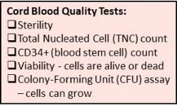
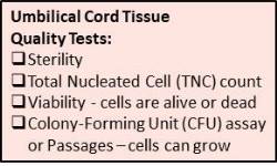
- Sterility: Every umbilical cord that arrives in the laboratory should be tested for sterility. Cells or tissue that may have bacterial, fungal or viral contamination are not suitable for therapeutic use.
- Cell Counts: If cells are isolated before cryopreservation, then cell counts can be reported. Unlike cord blood, pre-storage cell counts are not critical for MSC. Many therapies with MSC require that when the cells are taken out of storage they must be grown in culture to expand the cell count to a therapeutic dose13. Hence for MSC, cell viability and the ability to grow after thawing are much more important than counts of numbers of cells.
- Viability: Cell viability means the cells are alive, as demonstrated by their ability to grow and multiply successfully. The viability of the MSC is very important to know the true quality of the stored sample. Some banks routinely grow the cells through 2-4 passages before cryo-storage. Some banks prove viability by giving parents a photo of their cells growing in a culture dish. During cryo-preservation the thermal shock causes a drop in MSC viability, as low as 50%-25% of the pre-storage viability14. Hence it is important for the laboratory to conduct validation tests which demonstrate that the final product of their storage method is still viable.
- Colony Growth: The colony forming unit (CFU) assay is a test that confirms the growth of cells in culture, but ideally this test requires two weeks. Obviously it is not feasible to perform this test on individual samples prior to storage, but it should be performed as part of validation testing. The MSC from umbilical cord tissue have been found to have higher CFU-F frequency when compared to MSC from adult bone marrow15,16.
Cryopreservation: Because the processed umbilical cord tissue is stored at very low temperatures, a cryo-protectant is added to displace water out of cells so that they will not rupture as water expands during freezing. The cryo-preservation process generally begins with slow cooling at 1 degree per minute in a controlled rate freezer. The final storage temperature varies, with some banks using equipment at -800 Celsius and other banks using the same cryogenic nitrogen tanks that are used for cord blood which have temperature between -180 to -1960 Celsius. Regulating agencies such as the FDA, AABB, AATB, and FACT all require continuous monitoring of cryogenic freezers.
In summary, while parents already know that quality is important when choosing a family cord blood bank, parents need to know that they should also ask questions about the quality of the cord tissue storage method. If a bank doesn't have a validated method for processing cord tissue, the newborn's MSC may not be usable if they are needed for therapy. As part of their informed-decision making process, parents are encouraged to ask each bank about their quality testing of their cord tissue processing method.
Table of Umbilical Cord Tissue Processing Methods
Name | Description Cord Tissue Processing |
Intact Cord Tissue | Some companies store intact cord tissue as it is received. The tissue is cleaned and then stored intact by adding a cryo-protectant for future use. The MSC are not isolated from the cord. Studies find that it is difficult to extract MSC from previously frozen intact tissue17,18. |
Mechanical Methods | Some companies mince the cord tissue and cord lining membrane with scissors or scalpels and then store the small pieces. The pieces are immersed in a media or in the baby’s blood serum with a cryo-protectant. The sample can be stored in either bags or cryovials. This method is sometimes called “partial explants” or the “Freidman19 Approach”. It has been validated that isolated MSC can be retrieved from tissue stored in this manner20. |
Explants Methods | The explants method begins with mechanical mincing. Then the minced tissue is cultured in a container with suitable media and growth factors. As MSC grow out of the culture they adhere to the container surface and can be isolated for preservation. The explants processing method is the one most often used in clinical studies with MSC from umbilical cord tissue21,22, but it is also the most time consuming method. |
Digestion Methods | Digestion methods use enzymes such as trypsin, collagenase etc., to digest the tissue and its membrane lining. The enzymatic digestion destroys the extracellular meshwork and releases MSC and other cell types. Digestion is very efficient at releasing isolated MSC but studies suggest that the cell characteristics are somewhat different from the cells obtained without exposure to enzymes23,24. Some studies have looked at the trade-offs between mechanical versus enzymatic digestion25,26. |
TRT method | The method from Tissue Regeneration Therapeutics Inc. dissects the cord to extract the perivascular tissue, because this is the region where the MSC concentration is highest16,27. |
CliniMACS semi-automated methods | Semi-automatic methods use a mechanical device to isolate MSC from the matrix of the umbilical cord. The gentleMACSTM from Miltenyi Biotech GmBh “dissociates” a small segment of cord in a chamber. This device can be used with or without enzymes, but most published protocols employ enzymes28,29. |
AC:Px semi-automated method | The AC:Px from AuxoCell uses rotating blades to finely chop an entire umbilical cord in a closed chamber without any enzymes. This device yields both a minced tissue product and a cellular product30. |
 Dr. Asawari Bapat is originally from India, where she earned a medical degree with specialty in pathology. Dr. Bapat is experienced in the complete operational management for stem cell facilities. She is expert in regulatory compliance, having worked with AABB, FACT, JCI, CAP, US FDA, and the Ministry of Health for various nationalities. Dr. Bapat held a chair on the committee that developed the cord blood banking regulations adopted by the Indian Council of Medical Research (ICMR) and Drugs Controller General of India (DCGI). She has worked for the family cord blood banks Cordlife, LifeCell, Cryo-Save Arabia and Americord. Dr. Bapat has been based in Dubai for the past eight years, but does consulting on regulatory issues around the world. She is a member of the Advisory Panel to Parent’s Guide to Cord Blood Foundation. Dr. Bapat has working proficiency in eight languages.
References:
- Diseases Treated 2018; Available Online from Parent’s Guide to Cord Blood Foundation: ParentsGuideCordBlood.org/en/diseases (also available in the languages Spanish, Portuguese, Russian, Czech, Chinese)
- Perinatal Stem Cells, 2013; 2nd Edition, Cetrulo, KJ, Cetrulo, CL, Taghizadeh, RR, Editors, John Wiley & Sons Inc, Hoboken, NJ, USA.
- Cell Trials Data 2018; Available Online: CellTrials.org
- Can A, Celikkan FT, Cinar O. 2017; Cytotherapy. 19(12):1351–1382. Umbilical cord mesenchymal stromal cell transplantations: A systemic analysis of clinical trials
- Connick P, Kolappan M, Crawley C, et al. 2012; The Lancet Neurology. 11(2):150-156. Autologous mesenchymal stem cells for the treatment of secondary progressive multiple sclerosis: an open-label phase 2a proof-of-concept study
- Zhang J, Lv S, Liu X, Song B, Shi L. 2018; Gut Liver. 12(1):73-78. Umbilical Cord Mesenchymal Stem Cell Treatment for Crohn's Disease: A Randomized Controlled Clinical Trial.
- Natsumeda M, Florea V, Rieger AC, et al. 2017; J Am Coll Cardiol. 70(20):2504-2515. A Combination of Allogeneic Stem Cells Promotes Cardiac Regeneration.
- Ramireddy A, Brodt CR, Mendizabal AM, et al. 2017; Stem Cells Transl Med. 2017 May;6(5):1366-1372. Effects of Transendocardial Stem Cell Injection on Ventricular Proarrhythmia in Patients with Ischemic Cardiomyopathy: Results from the POSEIDON and TAC-HFT Trials.
- Lamo-Espinosa JM, Mora G, Blanco JF, et al. 2016; J Transl Med. 14(1):246. Intra-articular injection of two different doses of autologous bone marrow mesenchymal stem cells versus hyaluronic acid in the treatment of knee osteoarthritis: multicenter randomized controlled clinical trial (phase I/II).
- Soler R, Orozco l, Munar A, et al. 2016; The Knee. 23(4):647-654. Final results of a phase I–II trial using ex vivo expanded autologous Mesenchymal Stromal Cells for the treatment of osteoarthritis of the knee confirming safety and suggesting cartilage regeneration
- Iftimia-Mander A, Hourd P, Dainty R, Thomas RJ. 2013; Biopreserv Biobank. 11(5):291-8. Mesenchymal stem cell isolation from human umbilical cord tissue: understanding and minimizing variability in cell yield for process optimization.
- Couto PS. 2016; Transfusion. 56(6) ICBS1606 Poster Abstract DOI: 10.1111/trf.13686 Survey of How Cord Blood Banks Process Cord Tissue
- Centeno CJ, Al-Sayegh H, Bashir J, Goodyear D, Freeman MD. 2015; BMC Musculoskeletal Disorders. 16:258. A dose response analysis of a specific bone marrow concentrate treatment protocol for knee osteoarthritis.
- François M, Copland IB, Yuan S, Romieu-Mourez R, Waller EK, Galipeau J. 2012; Cytotherapy. 14(2):147-152. Cryopreserved mesenchymal stromal cells display impaired immunosuppressive properties as a result of heat-shock response and impaired interferon-γ licensing
- Lu LL, Liu YJ, Yang SG, et al. 2006; Haematologica, 91(8):1017-26. Isolation and characterization of human umbilical cord mesenchymal stem cells with hematopoiesis-supportive function and other potentials.
- Davies JE, Walker JT, Keating A. 2017; Stem Cells Transl Med. 6(7):1620–1630. Concise Review: Wharton's Jelly: The Rich, but Enigmatic, Source of Mesenchymal Stromal Cells
- Chatzistamatiou TK, Papassavas AC, Michalopoulos E, et al. 2014; Transfusion. 54(12):3108-20. Optimizing isolation culture and freezing methods to preserve Wharton's jelly's mesenchymal stem cell (MSC) properties: an MSC banking protocol validation for the Hellenic Cord Blood Bank.
- Badowski M, Muise A, Harris DT. 2014; Cytotherapy. 16(9):1313-21. Mixed effects of long-term frozen storage on cord tissue stem cells.
- Friedman R, Betancur M, Boissel L, Tuncer H, Cetrulo C, Klingemann H. 2007; Biol Blood Marrow Transpl. 13(12):1477-1486. Umbilical Cord Mesenchymal Stem Cells: Adjuvants for Human Cell Transplantation
- Skiles ML, Brown KS, Brown HL. 2015; Transfusion. 55:1A–7A ICBS1514 Poster Abstract Development of a Quantitative Measurement of Mesenchymal Stem Cells Explant Outgrowth from Umbilical Cord Tissue.
- Zhang Z, Lin H, Shi M, et al. 2012; J Gastroenterol Hepatol. 27(s2):112–120 . Human umbilical cord mesenchymal stem cells improve liver function and ascites in decompensated liver cirrhosis patients
- An Y-H 2016; Clin Transl Degener Dis. 1(1):9-16 Safety of intrathecal transplantation of human umbilical cord mesenchymal stem cells for treating hereditary cerebellar ataxia: study protocol for a randomized controlled trial
- Yoon JH, Roh EY, Shin S, et al. 2013; Biomed Res Int. 2013:428726. Comparison of explant-derived and enzymatic digestion-derived MSCs and the growth factors from Wharton's jelly.
- Taghizadeh RR, Cetrulo KC, Cetrulo CL. 2018; Cell Transplantation. DOI 10.1177/0963689717744787 Collagenase Impacts the Quantity and Quality of Native Mesenchymal Stem/Stromal Cells Derived during Processing of Umbilical Cord Tissue. (in publication, accepted 1 Nov. 2017).
- Maslova OO, Shuvalova NS, Sukhorada OM, et al. 2013; Dataset Papers in Biology 2013:370103. Heterogeneity of Umbilical Cords as a Source for Mesenchymal Stem Cells
- Emnett RJ, Kaul A, Babic A, et al. 2016; Stem Cells International. 2016:3274054 Evaluation of Tissue Homogenization to Support the Generation of GMP-Compliant Mesenchymal Stromal Cells from the Umbilical Cord
- Schugar RC, Chirieleison SM, Wescoe KE, et al. 2009; J Biomed Biotech. 2009:789526 High Harvest Yield, High Expansion, and Phenotype Stability of CD146 Mesenchymal Stromal Cells from Whole Primitive Human Umbilical Cord Tissue
- Fazzina R, Mariotti A, Procoli A, et al. 2015; Transfusion. 55(12):2864-73. A new standardized clinical-grade protocol for banking human umbilical cord tissue cells.
- Smith R, Pfeifer K, Petry F, Powell N, Delzeit J, Weiss ML. 2016; Stem Cells International. 2016:6810980 Standardizing Umbilical Cord Mesenchymal Stromal Cells for Translation to Clinical Use: Selection of GMP-Compliant Medium and a Simplified Isolation Method
- Auxocell Laboratories Inc. AC:Px 2017; Available Online: www.AuxoCell.com/tech.php.