Usted está aquí
Cord Blood Rescue After Cardiac Arrest
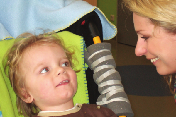
In this story, a child that underwent a prolonged cardiac arrest was resuscitated, but remained in a vegetative state because of the pause in blood flow to his brain. After an infusion of his own cord blood, he experienced a remarkable recovery over the next three years that has been published in the medical literature.
 The events that led to the cardiac arrest are every parent’s worst nightmare: A little boy in Germany, 2.5 years old, was perfectly normal before he became sick. He was admitted to the hospital after three days of vomiting. The boy underwent surgery and it was discovered that a loop of his intestine had twisted around itself, which created a bowel obstruction, which caused the non-stop vomiting. A portion of his intestine had died, causing a massive infection which spread into his blood. While he was in the hospital, the boy went into cardiac arrest. It took over 25 minutes and three rounds of defribrillation to restore his pulse and circulation. Very few children survive a cardiac arrest lasting more than 13 minutes.
Sadly, although the boy survived the cardiac arrest, he was diagnosed as being in a persistent vegetative state. Brain MRIs showed severe injury from ischemia (lack of oxygen). The doctors wrote that in a case like this, the “neurological prognosis of the patient… was ominous if not hopeless.” At best, such children only have minimal awareness of their surroundings at a follow up four years later.
The parents were desperate to try any therapy, so they contacted Vita 34, the cord blood bank where they had stored their son’s cord blood. Arrangements were made, consistent with German medical regulations, for the boy to receive an infusion of his own cord blood. This took place nine weeks after the cardiac arrest, and the child received followed up testing until 40 months later.
1 week follow up: When he was first admitted to rehabilitation, the boy cried and whimpered continuously. By one week after the cord blood infusion, he stopped whimpering and began to respond to acoustic stimuli.
2 month follow up: After two months, the patient was discharged from the rehabilitation center. During this time, his score of Gross Motor Function Measure (GMFM) had progressed from 0% to 23%, and his score on the Coma Remission Scale went from 33% to 92%. The boy could grasp, hold, bite, chew, and swallow a biscuit. Some of his eyesight was restored, he was smiling socially, and he could say “ma-ma.” Residual neurologic deficits included tetraparesis (muscle weakness in arms and legs), hypotonic trunk (also weak muscle tone), and other classic symptoms of cerebral palsy from the brain injury.
5 month follow up: At this visit the electroencephalogram (EEG) of brain electrical activity was normal! The boy made brief eye contact and was able to respond to questions by pointing at objects, but his expressive speech was very limited.
1 year follow up: Significant improvements were seen in fine motor control of the hands, social interaction, and cognition. He could sit unsupported but required support to stand. He still had spastic tetraparesis, particularly in the legs.
2 year follow up: At this time the patient was able to eat independently, move from prone position to free sitting, and crawl. He could walk with support. His vocabulary consisted of eight words, with slurred pronunciation. His fine motor skills had improved to such an extent that he could steer a remote controlled car.
40 months follow up: At the final follow up, his expressive speech had advanced to a vocabulary of 200 words and four-word sentences. He was pulling himself into standing position and walking independently in a gait trainer.
In conclusion, this “remarkable functional neuroregeneration is difficult to explain by intense active rehabilitation alone and suggests that autologous cord blood transplantation may be an additional and causative treatment of pediatric cerebral palsy after brain damage.”
Reference
Jensen A. & Hamelmann E. First Autologous Cell Therapy of Cerebral Palsy Caused by Hypoxic-Ischemic Brain Damage in a Child after Cardiac Arrest—Individual Treatment with Cord Blood. Case Reports in Transplantation 2013; 2013:951827. doi:10.1155/2013/951827


